
Microfluidics - new parasite diagnostic method for horses
Having many horses in a small area increases the risk of parasite infection, and parasites are also developing resistance to deworming agents. Diagnostics are time-consuming and outdated, and new methods are needed to limit the parasite burden and positively impact the horse's well-being.
Background
Horse parasites have developed resistance to deworming agents, which is a growing threat to horse health. As with antibiotics, deworming agents must be used restrictively in order to remain effective. Since 2007, selective deworming of horses has therefore been recommended, which in practice means that all horses are diagnosed and only those individuals that excrete a certain amount of parasite eggs are dewormed. A current problem is that infections with large bloodworms have increased, and that increase seems to have started after selective treatment was introduced. It is therefore important that horse owners choose the right parasite diagnostics in order to carry out the right treatment.
Current parasite diagnostics use sieving and centrifugation to separate parasite eggs from manure. The method is time-consuming. Apart from the resistance and health perspectives, it is also not cost-effective to use unnecessary treatment on entire herds instead of performing simple diagnostics. SLU and LTH are collaborating to develop a new diagnostic tool for parasitic infections in horses. The new method is based on advanced microfluidics combined with a mobile phone camera. The diagnostics can be performed on site by animal owners in less than an hour.
Results
The aim of this pilot project was to investigate whether microfluidic technology could be used to diagnose parasitic diseases in horses. The long-term goal is for this technology to provide a more sensitive and selective diagnosis than current methods for parasite diagnosis.
The difficulty with fecal samples is that they are very complex and therefore require several separation steps in this process. In simplified terms, the process can be described using the following example steps:
- Plant parts larger than the parasite eggs are removed. The parasite eggs are concentrated.
- Sorting and collection of parasite eggs.
- Identification and counting and/or separation of eggs.
We have started with step 1, where particles are sorted by size using Deterministic Lateral Displacement (DLD). Microfluidics will be able to be combined with integrated microscopy, based on the camera found in most mobile phones. It will be easy for the user. The goal is to take a stool sample, place it on the device, and add a saline solution. The solution is then pumped into the microfluidics and analyzed via an add-on that can be connected to a mobile phone, which reads the result.
We also believe that the new parasite diagnostics using microfluidics technology will be both cheaper and easier to use.
Participants in this project:
- Eva Tydén, SLU
- Jonas Tegenfeldt, LTH.
- Jason Beech, LTH.
Method of analysis
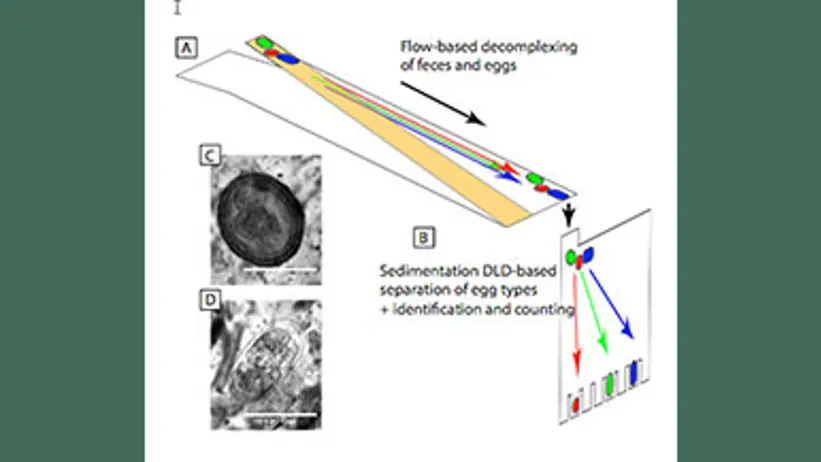
Figure 1: Schematic diagram of egg separation from feces. (A) Flow-based decomplexation of feces. (B) DLD based on sedimentation for sorting different types of eggs. (C) Hookworm eggs (D) Bloodworm eggs.
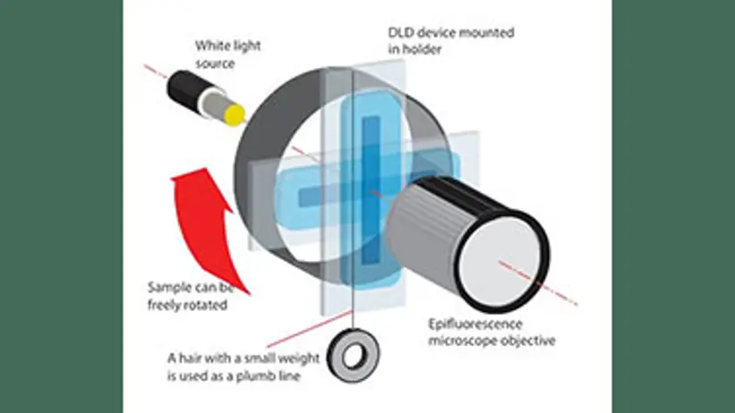
Figure 2: Experimental sketch of the sedimentation-driven DLD device for particle sorting.
Funding
The project, which began in 2018 and lasted for one year, was funded by SLU Future One Health, which supports forward-looking interdisciplinary research within One Health – good health and welfare for humans and animals in sustainable ecosystems.
Contact
-
PersonEva Tyden, ResearcherHBIO, Division of Food Safety, Infection Biology, Pharmacology and Toxicology (LIFT)