
The Eye’s Fragile Window: Research for rapid and reliable treatment of corneal ulcers in animals
The cornea is the eye’s window to the world and its first line of defence. Ingeniously built but highly vulnerable, it requires swift and accurate treatment when injured. Researchers in Sweden are now working to improve treatment of corneal ulcers in our most cherished animal species.
The cornea – a transparent shield between the eye and the world
Much like a car windscreen, the cornea serves two vital purposes: it allows clear vision through its transparent structure, while simultaneously protecting the valuable and delicate tissues behind it. The eye is also assisted by tear fluid, which – like windshield washer fluid – is constantly present and can rinse away debris when visibility is compromised. And of course, there are the eyelids, blinking like windscreen wipers to sweep away dust, eyelashes and other unwanted intruders.
But the transparent window that the cornea forms between the eye and the world only functions as intended as long as it remains intact. In a collision, the windscreen may crack – and what happens then?
When a small injury becomes a big problem
There are many reasons why corneal wounds and ulcers may occur. In dogs, tear production or tear film quality may be inadequate, failing to keep the cornea sufficiently lubricated and protected against dust, eyelashes and debris entering the eye. In both dogs and cats, a claw during a fight can easily perforate the cornea.
And suddenly, the very feature that makes the cornea so effective – its transparency – becomes its greatest weakness.

Smart yet fragile – transparency at a cost
Because the cornea is transparent, it contains no blood vessels.
Elsewhere in the body, the immune system is an extensive and efficient network. Through interconnected blood vessels, white blood cells can be rapidly transported to wherever they are needed. This allows bacteria and other foreign agents to be stopped at the breach that a wound represents, while the healing process begins.
Instead, the body has to start building new blood vessels into the cornea so that immune cells can reach the injured area of the eye more effectively.
Understanding this, it becomes clear why veterinary medicine strives to assist the healing of corneal ulcers – and why this research project was initiated.
Because it is not only about treating quickly, but about treating precisely.
Precise treatments reduce the risk of antibiotic resistance
To prevent bacteria from invading the eye, corneal injuries are treated as soon as they are detected – most often with antibiotics. But treatment must be targeted and appropriate.
Antibiotics are medicines used to treat bacterial infections.
Antibiotic resistance develops when bacteria evolve mechanisms that render antibiotics less effective, or completely ineffective. This is a serious and growing public health problem, both in Sweden and globally.
To avoid contributing to this problem, the research group aims to identify which bacteria most commonly infect corneal ulcers in animals in Sweden. This knowledge will help Swedish veterinarians make informed decisions when treating suspected infections and choose antibiotics with the highest likelihood of targeting only the bacteria actually present in the damaged cornea.
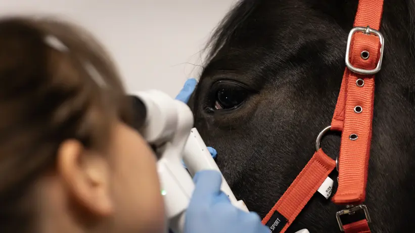
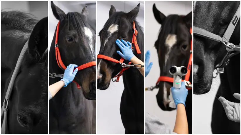
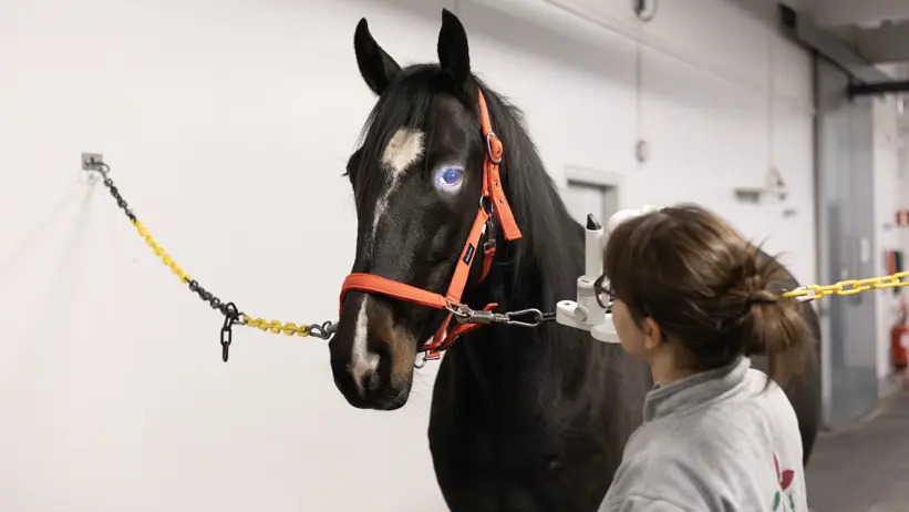
– Treatment of corneal injuries often consists of eye drops, eye drops – and yet more eye drops. So it really matters that you are using the right ones, says Hanna Holmqvist, doctoral student in the project.
One foot in the lab, the other at the clinic
When a deeper or more complicated corneal ulcer is left to heal on its own, the eye gradually forms new blood vessels that grow from the edge of the cornea towards the wound – like fine threads creeping across an otherwise transparent surface
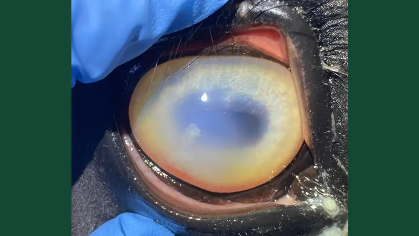
In the best-case scenario, these blood vessels regress once the injury has healed and the visual field clears again.
Research with a clear goal: safer, sharper treatments
Until 2029, Hanna Holmqvist will devote her full working time to this project, aiming to improve the safety and precision of treatments for corneal ulcers in animals.
With a veterinary degree under her belt, she is able to contribute both by assisting at the eye clinic with sampling and patients, and in the lab with examining and analyzing.
Already within the first months of the project, more than 200 samples from horses, dogs, cats and rabbits have been collected – a strong start to the four-year study.
- Investigate which factors influence the healing of corneal ulcers.
- Map the bacterial species most commonly associated with infected corneal ulcers in horses, dogs, cats and rabbits in Sweden.
- Evaluate which types of antibiotic treatment are effective against specific bacterial pathogens, and, based on this knowledge, assess whether current Swedish veterinary treatment guidelines should be revised.
– Although a substantial body of international research already exists, differences in climate, animal breeds, and Sweden’s comparatively strict antibiotic policy mean that international recommendations are not necessarily optimal under Swedish conditions. This may ultimately influence which treatments are most appropriate to use here, explains the doctoral student.
In addition, the research group aims to compare different diagnostic sampling methods, to ensure that test results are consistent regardless of which method is used.
– This includes both sampling with a special type of cotton swab for bacterial culture and sampling with a cytology brush for cellular analysis, Hanna Holmqvist explains, and continues:
– The latter is much faster, as the brush can be examined directly under the microscope. But does it provide the same information as a swab that is cultured in the laboratory, allowing bacteria time to grow to detectable levels? That is what we want to find out.
Take home advice to readers and animal owners: “All eyes need help – and they need it fast”
Key advice from veterinarian and doctoral student Hanna Holmqvist:
- The eye is a delicate structure. If something seems wrong, seek medical or veterinary attention immediately – whether it concerns you or your animal.
- A wound to the eye is not comparable to a wound elsewhere in the body. Because the cornea lacks blood vessels in order to remain transparent, the body’s immune response is much slower here. If an infection develops, there is a real risk of permanent damage and impaired vision.

About the project
- Officially titled: “Infectious ulcerative keratitis in horses and companion animals in a Swedish context; prevalence of bacteria and fungi, resistance patterns and prognosis of healing”
- Expected to run during the period 2025-2029
- Funding bodies include the Horse Research Foundation, The Greater Stockholm Veterinary Care Foundation and the Agria and SKK Research Fund.
- The doctoral student in the project is Hanna Holmqvist, together with a research group consisting of:
- Ingrid Hansson, principal supervisor and professor of bacteriology at the Swedish University of Agricultural Sciences (SLU).
- Lena Ström, veterinarian, European eye specialist, and researcher at SLU.
- Katarina Nostell, veterinarian and associate professor of internal medicine at SLU.
- Oskar Nilsson, antibiotics expert and laboratory veterinarian at the Swedish National Veterinary Institute (SVA).
- Plus two clinically active veterinarians undergoing training to become Swedish eye specialists: Sara Littorin and Kristian Poulsen.
The project can be broadly divided into the following parts:
- The project will begin during the first two years with the collection and analysis of samples from horses, dogs, cats, and rabbits.
- The latter two years will then be devoted to mapping the collected and analyzed material, focusing on the following questions:
- Which bacteria are found in each animal species in Sweden?
- Is it possible to identify which types of antibiotics the above bacteria are sensitive to (known as resistance patterns)?
- Is it possible to equate different diagnostic methods?
As a complement to the study, the research team will also work to increase understanding of why some corneal ulcers take longer to heal, or do not heal at all.
Contact
-
PersonHanna Holmqvist, PhD studentHBIO, Bacteriology, Virology, Food Safety and Veterinary Public Health